MRI Equipment and Resources
The heart of the CABIN is a Siemens MAGNETOM Prisma 3T whole-body scanner that includes:
- A gradient system of maximum amplitude of XR 80 mT/m @ 200 T/m/s, and high order shim
- 64-channel and 20-channel head and neck coils (capable of parallel imaging using SENSE/GRAPPA), with dedicated brain pulse sequences allowing many types of research applications.
- Simultaneous multiple slice (SMS) techniques for acquisition of BOLD and DTI results in time saving, higher spatial/temporal resolution, and/or combinations of each.
- Spine Dot Engine provides optimized cervical, thoracic, and lumbar spine imaging.
- Full assortment of other coils including extremity and body coils as well as small animal custom coils, which allow a wide range of other applications, such as musculoskeletal, cardiac, liver and animal imaging.
- A research agreement with Siemens Healthineers to provide access to pulse sequence programming platform, access to on-going work-in-progress (WIP) software packages, and exchange agreements with researchers in other institutes.
Additional equipment is provided for stimulus presentation and subject response and physiological monitoring:
- Computer interfaced with the scanner for synchronization, video display (in room BoldScreen 32, Cambridge Research Systems, with refresh rate up to 120Hz), audio equipment (SereneSound audio system, Resonance Technology Inc. 30dB gradient noise attenuation, 40Hz to 40kHz frequency response, active noise cancellation patient microphone), microphone and response boxes (fORP 904 Subject Response Package, Cambridge Research Systems) to present stimuli and record subject responses
- Standard software packages for stimulus display and response recording (e.g., Presentation, E-Prime, Psychophysics Toolbox and MATLAB).
- FIRMM, NOUS Imaging Inc. system provides real-time monitoring and biofeedback technology that addresses the problem of patient motion during brain MR.
- BIOPAC system (BIOPAC Systems, Inc.) for recording of physiological signals in the MRI environment such as: Electrocardiogram (ECG), Temperature, Respiration, Pulse Wave, ETCO2 and O2 Gas Recording and Analysis, Pulse Oximeter (SpO2). The accompanying AcqKnowledge software allows physiological data viewing, measuring, transformation, and analysis.
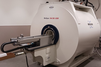 The 9.4T/30 cm bore Bruker BioSpec MRI system is a zero Helium boil-off Ultra Shield Refrigerated (USR) magnet for unsurpassed sensitivity in small animal MRI studies.
- State of the art Paravision 360 software provides a host of over 100 validated and ready-to-use in-vivo protocols and scan programs for mice and rats with integrated examination guide for optimal scanning navigation and reproducibility.
- Paravision 360: Dynamic shimming capability, B1 optimization and mapping, Fat-water separation imaging, and Fat chemical shift corrected images. Provides extensive reconstruction, viewing and analysis functions, ranging from angles and annotation to surface rendering to underlay/overlay, to zooming/panning to automatic 3D image fusion.
- The 9.4T MRI system is equipped with two gradient/shim systems (main B-GA20S HP and an insert B-GA12S) that enable 660 mT/m gradient strength and 4570 T/m/s slew rate for optimum gradient/shim performance for small animal MR imaging.
- Wide range of RF coils to meet the varying needs of MRI research.
- Mouse/Rat Volume Coil (112/86 mm)
- Rat Body Tx/Rx Volume Coil (112/72 mm)
- Guinea pigs or rabbits Volume Coil (198/154 mm)
- Rat Head & Mouse Body Tx/Rx Volume Coil - 40mm
- Mouse Head Tx/Rx Volume Coil - 23mm
- Mouse Brain Array Coil
- Rat Brain Array Coil
- Mouse Heart Array Coil
- Mouse Brain Opto-Genetics Array Coil
- Multi-Purpose Planar Surface Coil – 10/20/30 mm
- 1H/31P Tx/Rx surface coil- 10 mm
- Arterial Spin Labeling (ASL) coil for Rat Head
- CryoProbe Mouse Brain Array Coil
- PET Tx/Rx Volume Coil (72 mm)
- 1H CryoProbe Mouse Brain Array Coil: Cryogenically cooled RF coils (20 - 30 K) which provides a remarkable SNR gain compared to RF coils operated at room temperature. This can be used for in-vivo imaging with higher resolution up to 20 µm and for new applications that are not feasible with room temperature coils.
- Autopac: Motor driven automatic animal positioning system for cradles.
- Small Animal Monitoring System:
- Basic Life Monitoring and MRI gating Unit (SA Instruments, Inc., model 1030):
- Display and analysis of physiological (ECG and respiratory) signals.
- Body temperature measurement and supervision
- ECG or respiratory synchronized MRI gating
- Small Animal Anesthesia System with vaporizer and either compressed room air or oxygen concentrator
- Thermo Scientific SC100 Animal Temperature Conditioning System: Water based heating unit to condition the body temperature of (anesthetized) rodents via warming blankets or integrated water hoses in animal beds.
- Basic Life Monitoring and MRI gating Unit (SA Instruments, Inc., model 1030):
- PET Insert Si 198: For simultaneous PET and MR imaging of mice and rats. This allows researchers to gain unique insight on molecular and functional processes as well as a multitude of macroscopic and chemical information within a living system at the same time.
- Other Services:
- Trained MR staff to assist with imaging and animal/sample preparation.
- Harvard Apparatus PHD 2000 Infusion pump
- Animal preparation rooms equipped with amenities such as CRC-55tW Dose Calibrator with Well Counter, pipettes, surgery tools, recovery area, animal holding racks, snorkel exhaust system and vaporizer with compressed air or oxygen tanks.
The EyeLink 1000 Plus, integrated within our MRI scanner, allows for precise and reliable eye-tracking during neuroimaging studies. This system captures a wide range of eye-tracking data, including saccades, fixations, blinks, and pupil diameter, with exceptional accuracy and speed. These data provide real-time insights into eye movement patterns, visual attention, cognitive functions, and emotional responses. At the University of Rochester Center for Advanced Brain Imaging & Neurophysiology, this technology is utilized in various cutting-edge research areas, including virtual reality studies, attention research, resting-state visual attention, and more. The EyeLink 1000 Plus is fully compatible with the MRI environment, ensuring high-quality data acquisition without interference. This advanced setup supports a broad spectrum of research applications, from basic neuroscience to clinical studies, offering invaluable insights into the interplay between eye movements and brain activity.