News
Heard of the Microbiome? Meet the MicroRNAome.
Tuesday, October 31, 2017
By Susanne Pallo
A University of Rochester Medical Center researcher is part of a team that is working toward characterizing where a certain type of RNA, called microRNA, is expressed in human cells. In a recent study, published in Genome Research, the team made a giant set of data about these RNA available to the public to guide research and foster the development of new therapies.
RNA, the close cousin of DNA, comes in many flavors - each with a specific role. MicroRNA (miRNA) help regulate which proteins are produced in a cell and how much. Fiddling with the level of proteins can have subtle or large impacts on that cell's activity and can even cause disease.
Several studies have linked miRNAs to diseases, either as the cause or simply a marker. While some of these links could lead to new treatments, others may just be red herrings.
"We've showed previously that when somebody claims a certain miRNA is a marker of a disease, at times it's just a marker of increased inflammation, which is a side effect of a lot of diseases," said Matthew N. McCall, Ph.D., assistant professor of Biostatistics and Biomedical Genetics at URMC and an author on the study.
According to McCall and study leader Marc K. Halushka, M.D., Ph.D., associate professor of Pathology and director of Oncology Tissue Services at Johns Hopkins University School of Medicine, before you can understand what miRNAs are doing, you need to know where they are.
And why is that important?
Within a given tissue in your body, there could be thousands of different cells types, serving different purposes. When measuring RNA in a tissue biopsy, you will get a mixture of RNA from all of these different cell types, whether or not they are relevant to the disease you are interested in.
For their study, Halushka's team pulled together all that was known about miRNA in human cells and conducted experiments to fill the gaps. Though it wasn't their specific intent, the researchers found that many miRNAs originally thought to be expressed in all or many cell types actually are not. Rather, they are expressed in cells that get into all or most tissues, like blood or inflammatory cells, and could be mistakenly associated with a disease.
All of the data from Halushka's study are available in an online database and in the University of California, Santa Cruz Genome Browser and will be updated regularly. Having access to this data should save researchers a lot of time and help weed out some those aforementioned red herrings. With just a few clicks, researchers can find all of the cells types that express a specific miRNA or all of the miRNAs expressed in a specific cell type.
“Bubbles” Boost Search for Treatment to Aid Head and Neck Cancer Patients
Wednesday, October 25, 2017

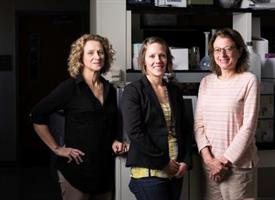
Catherine Ovitt, Danielle Benoit, and Lisa DeLouise
A scientific team at the University of Rochester is using innovative technology to discover preventative treatments for salivary gland radiation damage typical for head and neck cancer patients—and recently received a $3.8 million National Institutes of Health grant to support their investigation.
Cancer patients can lose salivary gland function during treatment for head and neck tumors. The irreversible damage, which prevents patients from producing saliva, often results in permanent dry mouth and makes it difficult to eat, speak, and swallow. The team will develop salivary gland tissues using a unique chip technology called "microbubbles," which are tiny spherical wells or bubbles that can hold cells.
The use of the microbubble platform is based on several years of salivary gland research, led by Catherine E. Ovitt, Ph.D., associate professor of Biomedical Genetics, a member of the UR Center for Oral Biology, and an expert in the repair and regeneration of salivary glands, and Danielle Benoit, Ph.D., associate professor of Biomedical Engineering and an expert in drug delivery systems and hydrogel platforms for tissue engineering approaches. Together with Lisa A. DeLouise, Ph.D., associate professor of Dermatology and Biomedical Engineering, who developed and received several patents for the microbubble concept, the scientists are working as co-principal investigators on the NIH project.
Their goal is to find drugs that could be given to patients prior to radiation treatment that would prevent damage to the glands.
"Dr. Ovitt and I have shown through years of investigation that being able to develop functional salivary gland tissue for testing is the key to solving this problem," Benoit said. "So, it's microbubbles to the rescue."
Expanding cells and tissue outside of the body is elusive. In this case the process involves taking salivary gland cells that have been removed from humans undergoing surgery, expanding the cells, and studying their reaction to various drugs.
A major problem, however, starts to occur as soon as the tissue is removed from the body and isolated: Cells immediately begin to lose their natural function. In the body, cells send signals and secrete proteins that are essential for their survival. In a culture plate in a laboratory, however, these signals and proteins are diluted and dispersed, making the cells no longer viable.
DeLouise's technology at first glance looks similar to a cell culture petri dish, a round piece of silicone about the size of the large cookie. But within the dish are an arrangement of thousands of tiny round "micro-wells," each one comprising a minuscule compartment for cell growth and tissue formation. The unique shape of each microbubble creates a niche that concentrates the cells, allowing them to proliferate and form salivary gland units.
The microbubbles come in different sizes, and the beauty of the technology is that scientists can grow cells in thousands of bubbles at one time. DeLouise can make dishes the size of a dime that include more than 5,000 microbubbles. In addition, Benoit's lab has produced hydrogel materials that can be placed inside each microbubble that further allow the cell to maintain its structure and function.
If the team can successfully grow human salivary gland cells in the microbubbles, they say, they will also be able to rapidly test thousands of existing Food and Drug Administration-approved drugs on the salivary tissue using the microbubble technology.
"Only one treatment is currently available for radioprotection but it comes with many side effects, so most patients discontinue it," Ovitt said. "There is a great need for additional ways to either cure or prevent this debilitating condition."
The team is collaborating with Shawn D. Newlands, M.D., Ph.D., M.B.A., chair of the Department of Otolaryngology and member of the Wilmot Cancer Institute's head and neck oncology team, to collect salivary tissue from consenting patients undergoing salivary gland surgery. Salivary gland cells are isolated from these tissues for seeding into microbubbles for the investigation. Additionally, Paul Dunman, Ph.D., associate professor of Microbiology and Immunology, will provide high-throughput drug-screening expertise during the second phase of the project, which is contingent upon successful development of the human gland chips.
GDSC Team supports the 2017 Wilmot Cancer Warrior Walk
Sunday, September 10, 2017
Several GDSC students and faculty attended the 5th Wilmot Cancer Warrior Walk this Sunday. Showing off our colors in form of this year's new GDSC T-shirts, the Team participated very successfully in the 10K, 5K and 1M events. Adam Cornwell, Andrew Albee, Xiaolu Wei, Fanju Meng, Justine Melo and Dalia Ghoneim were our "Runing Warriors" for the 10k and 5k events, with Andrew Allbee finishing overall 6th (44:05 min, 3rd 20-29yr old) and Adam Cornwell 7th (44:40 min, 1st 30-39yr old) in the 10k. Dalia Ghoneim ran the 5k and was the 3rd overall female finisher, and was 2nd in her age category (30-39 years). Her time was 23:33. Congratulations to all! The team was rounded out by Dashiell Na, Shen Zhou and and Anne Roskowski. Led by the scientific director of the Wilmot Cancer Center, Hucky Land, faculty also attended in force, including the whole Samuelson family, cancer biologist Mark Noble and the Pröschel's. Runners and walkers alike enjoyed a beautiful sunny day out and the positively uplifting company of cancer fighters and survivors! See you all again in 2018!
Facebook Post
GDSC Picnic Kicks-Off the 2017-2018 Academic Year
Thursday, September 7, 2017
Braving at times inclement weather, Graduates students and Faculty of the Genetics, Development and Stem Cell program gathered at he Genessee Valley Park hawthorne lodge for food, fun and festivities. After a week of rain and clouds from the outer bands of Hurricane Harvey, we caught a break and ended the day with sunshine! A fitting start to the new academic year and a warm welcome to Anne Roskowski, a new graduate student in the GDSC program.
Doctors Might FINALLY Be Able to Tell If Your Infection Is Bacterial or Viral
Monday, August 14, 2017
If you head to your doctor with a fever and cough or other signs you're getting sick, there's a good chance your doctor won't know what's behind it. It's hard to test for certain diseases, like bacterial pneumonia.
When patients come in with respiratory viruses like the common cold and sinus infections, doctors often send them home with an antibiotic prescription. The problem is, antibiotics only target bacteria and won't do anything to fight a virus. Still, about 30 percent of antibiotics prescriptions are for viral infections, according to the CDC. (Make sure to ask these essential questions before taking antibiotics.)
Not only is an antibiotic completely unhelpful against a virus, but it could have major consequences. Antibiotics kill most of the bacteria behind your infection—but not all of them. The bacteria that are strong enough to survive will multiply to create more bacteria that are resistant to treatment, too. Over time, this means there will be more resistant bacteria than ones that antibiotics can kill, so the infections will be harder to treat.
And if you take antibiotics when you don't need to, or use antibacterial soap, you're speeding that process along. Eventually, diseases that used to be easy to treat with antibiotics could become dangerous again.
Luckily, researchers might have found a way to tell the difference between bacterial and viral infections, so you won't get a useless antibiotic. In a study in the journal Scientific Reports, researchers took blood from 94 patients who had lower respiratory tract infections. Lab tests found genetic markers in the blood that could correctly figure out if an infection was viral or bacterial 80 to 90 percent of the time. (Find out how genetic tests could help you lose weight, too.)
Because the sample size was so small, more tests will be needed before doctors can start using this diagnosis method in their offices. But if it does take off, it will be a lot easier than testing for specific diseases.
"Our genes react differently to a virus than they do to bacteria," says study co-author Thomas Mariani, PhD, pediatrics, environmental medicine, and biomedical genetics professor at University of Rochester Medical Center, in a statement. "Rather than trying to detect the specific organism that's making an individual sick, we're using genetic data to help us determine what's affecting the patient and when an antibiotic is appropriate or not."
In the meantime, if you think you're not feeling well, use these natural remedies for cold and flu.
Congratulations Margaret!
Wednesday, July 26, 2017
Margaret Hill
On Monday PhD candidate Margaret Hill presented her work investigating intrahepatic cholangiocarcinoma (iCCA), a form of liver cancer which morphologically resembles the biliary tract. Margaret completed her work under guidance of Dr. Aram Hezel. Her work helps us to understand the interplay between chronic liver injury, a common risk factor for this cancer and the cell of origin as she proved that hepatocytes, as opposed to biliary cells, may serve as a cell of origin for this cancer. Further investigation into important pathways known to be activated in biliary-derived iCCA showed hepatocyte-derived iCCA similarly up-regulates the Wnt and Notch pathways and thus could be targeted for treatment. Margaret went on to probe the importance of MCL-1, the most commonly amplified gene in iCCA, and identified a genetic subset of iCCA cancers which appear to depend on MCL-1 expression. Together, Margaret's work may have important therapeutic implications for iCCA. Well done Margaret and congratulations to Aram!
Hidden Herpes Virus May Play Key Role in MS, Other Brain Disorders
Monday, July 10, 2017
The ubiquitous human herpesvirus 6 (HHV-6) may play a critical role in impeding the brain’s ability to repair itself in diseases like multiple sclerosis. The findings, which appear in the journal Scientific Reports, may help explain the differences in severity in symptoms that many people with the disease experience.
“While latent HHV-6 – which can be found in cells throughout the brain – has been associated with demyelinating disorders like multiple sclerosis it has not been clear what role, if any, it plays in these diseases,” said Margot Mayer-Proschel, Ph.D., an associate professor at the University of Rochester Medical Center Department of Biomedical Genetics and co-author of the study. “These findings show that, while in the process of hiding from the immune system, the virus produces a protein that has the potential to impair the normal ability of cells in the brain to repair damaged myelin.”
Stem Cells May Be the Key to Staying Strong in Old Age
Tuesday, June 13, 2017
University of Rochester Medical Center researchers have discovered that loss of muscle stem cells is the main driving force behind muscle decline in old age in mice. Their finding challenges the current prevailing theory that age-related muscle decline is primarily caused by loss of motor neurons. Study authors hope to develop a drug or therapy that can slow muscle stem cell loss and muscle decline in the future.
Video of 3 Minute Thesis Event
Thursday, June 8, 2017
We have the video of the full event with all presentations fully captions and with the slides running in time with the videos.
3MT Presenters, Programs & Topics
Thesis presentations in order
- Stephanie Carpenter (Chemistry) - Solving the Mystery of Iron Chemistry
- Sarah Catheline (Pathways of Human Disease) - Inhibiting Inflammaging to Treat Osteoarthritis(OA)
- Scott Friedland (Genetics, Development & Stem Cells) - Pancreatic Cancer and the Tale of the Broken Librarian
- Claire McCarthy (Toxicology) - Investigating the Toxicological Effects of Dung Biomass Smoke Exposure
- Taylor Moon (Immunology, Microbiology and Virology) - The New Epidemic
- Thuy-Vy Nguyen (Social-Personality Psychology) - Solitude *Winner*
- Manisha Taya (Cellular & Molecular Pharmacology and Physiology) - Understanding Lymphangioleiomyomatosis (LAM): The “Other” Steroid-Dependent Cancer From Bed-Side to Bench and Back Again
- Janelle Veazey (Immunology, Microbiology and Virology) - Role of Protein Kinase D in Epithelial Cells During Respiratory Infection
Scott Friedland takes 2nd place in the Three Minute Thesis (3MT) competition
Monday, May 15, 2017
On May 11th, 2017, Scott Friedland took 2nd place in the Three Minute Thesis (3MT) competition with his talk entitled, “Pancreatic Cancer and the Tale of the Broken Librarian. 3MT, created at The University of Queensland in Australia, is an effort to bring awareness to research and scientific communication, in which competitors have 3 minutes to get across the thrust of their thesis to a general audience. Scott is an MD/PhD student currently working in the lab of Dr. Aram Hezel in the Genetics, Development, and Stem Cells program. His research focuses on defining the role of ARID1A and the SWI/SNF complex in pancreatic cancer and development.
29th Annual Genetics Day Symposium
Thursday, May 4, 2017
This year's Genetics Day provided another opportunity to celebrate the impact of Genetics on science and medicine. The program consisted of four short talks by U of R scientists, a poster session and the Keynote event, the Fred Sherman Lecture, delivered this year by Dr. David Sabatini from the Whitehead Institute for Biomedical Research at MIT. Dr. Sabatini talked about “Growth Regulation by the mTOR Pathway”.
This year’s Genetics Day Poster Session included posters from research laboratories across the University. Three graduate students and one post-doctoral associate were awarded poster prizes:

Andrew Allbee – Biteau Lab
dLMX1A is Required for Drosophila Ovary Stem Cell-Niche Unit Establishment
Amber Cutter – Hayes Lab
Molecular Characterization of Nucleosome Recognition by Linker Histone H1.0
Browyn Lucas – Maquat Lab
Evidence for Convergent Evolution of Sines for Staufen-Mediated Control of MRNA Decay
Janet Lighthouse – Small Lab
Expression Profiling Reveals the Cardioprototective Role of Metallothioneins in Exercise
Genetics Day has been a long-standing tradition at the University of Rochester and more recently includes the Fred Sherman lecture in memory of Fred Sherman a renowned biochemist and geneticist, who led international efforts to establish the yeast Saccharomyces cerevisiae as the premier genetic eukaryotic model system. The lecture is made possible by a generous fund endowed by Fred Sherman's wife, Elena Rustchenko-Bulgac, herself a research professor at the URMC.
GDSC Graduate Justin Komisarof successfully defends Friday April 28
Tuesday, May 2, 2017
MD/PhD candidate Justin Komisarof presented his work on the hunt for a common cancer gene signature. Building on prior work by the Land and McMurray labs, Justin used bioinformatic approaches to search for the presence of a ‘cooperation response gene’ (CRG) cancer gene expression signature a broad panel of different tumors. Using microarray gene expression datasets from GEO in colon, pancreatic, prostate, and head and neck cancers, Justin found a core set of consistently disregulated CRGs. A more in-depth study of genotype-phenotype correlation in colon cancer samples revealed that this CRG dysregulation did not track with p53 or Ras mutations, suggesting that CRG dysregulation occurs in multiple human cancers, and that the CRG expression pattern is independent of mutational status and may emerges as a function of the malignant state. Justin also applied this CRG-based analysis to prostate cancer samples and identified a core set of CRGs that when combined with clinical staging predicts recurrent outcomes with greater than 80% accuracy – a highly significant improvement over current methods. Justin’s work provides an important step forward for both diagnostic and future therapeutic approaches.
A four gene signature predictive of recurrent prostate cancer. Komisarof JMcCall M, Newman L, Bshara W, Mohler JL, Morrison C, Land H. Oncotarget. 2017 Jan 10;8(2):3430-3440.
GDSC Students attend the March for Science
Tuesday, April 25, 2017
Students from the Genetics Program attended The Rochester March for Science on Saturday April 22
Fanju Meng (Biteau Lab), Sreejith (Biteau Lab), Emily Wexler (Portman Lab),
Sebastian Rojas Villa (Biteau Lab), Robert Hoff (Bohmann Lab), Andrew Allbee (Biteau Lab)
Michael John Beltejar awarded CTSI Pilot Trainee Grant
Thursday, April 13, 2017
Michael John Beltejar, a 4th year student in the Genetics, Development, and Stem Cells PhD program with a research focus in Genetic contributions to bone strength has been awarded CTSI Pilot Training Grant.
CTSI Pilot Trainee Grant RFA
A major priority for the CTSI is the active support of research collaborations via cross-disciplinary collaboration, and the support of research that addresses significant problems related to population health. Thus, applications directly addressing these areas are strongly encouraged. Trainee awards help awardees obtain the most prestigious fellowship possible following the project. The project should be part of a long-term plan to become an independent investigator. The award provides a maximum of $25,000 for a period of one year.
Project Description
Osteoporosis remains a significant concern. By 2025, osteoporotic fractures are expected to increase to 3 million with an estimated cost of $25.3 billion. Morbidity and mortality increases following all major fractures in patients over 55 years of age. It is widely understood that compromised bone strength is the underlying pathophysiology of osteoporosis. Currently, bone mineral density (BMD), is the basis for diagnosis and the main target for osteoporosis therapy. BMD is an important clinical tool, but explains only 55 % of fractures, leaving the needs of 45% of patients unaddressed. Fundamentally, bone strength has two interconnected but distinct components: quantity and quality. While BMD reflects quantity, bone quality (BQ) reflects morphologic and compositional properties. However, developing therapies based on BQ is limited by two major gaps in knowledge: 1) Which genes regulate BQ and 2) Does estrogen deficiency modify the genetic regulation of BQ? Therefore, the objective of the work proposed is to construct a genetic network to identify candidate genes regulating bone composition(SA1) and determine if estrogen deficiency interacts with these genes(SA2).
The rationale for the proposed work is that bridging these gaps in knowledge is a crucial first step in the identification of novel therapeutics. The results produced from SA1 will inform ongoing research in the lab--in particular, a genome-wide association study identifying locations in the genome that are responsible for bone quality. Secondly, the results of SA2 have a strong potential to unlock research on multiple levels osteoporosis management. Identification of novel genes could be used as a new biomarker to identify at risk individuals earlier. Downstream activity of these genes could become additional metrics to monitor disease progression. Finally, these genes become putative targets for novel therapies. The research in this application is innovative because it is a significant departure which shifts focus from BMD to BQ as an equally important contributor of fracture resistance.