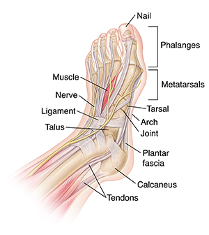
Anatomy of the Foot
The foot is one of the most complex parts of the body. It consists of 28 bones connected by many joints, muscles, tendons, and ligaments. The foot is prone to many types of injuries. Foot pain and problems can cause pain and inflammation, limiting movement.
-
Muscles contract and relax to move the foot.
-
Tendons are tough fibers that connect muscles to bones.
-
Ligaments are fibrous strands that connect bones.
-
Nerves travel throughout the foot, providing feeling.
-
Nails protect the tips of the toes.
-
Phalanges are the toe bones.
-
Metatarsals are the bones between the toes and the ankle bones.
-
Tarsals are bones of the rear foot (hindfoot) or middle foot (midfoot).
-
The talus is one of the ankle bones.
-
The calcaneus is the heel bone.
-
The arch is formed by bones and held in place with ligaments.
-
Joints are the meeting points between two bones. They are lined with cartilage. Cartilage is smooth tissue that allows joints to move easily.
-
The plantar fascia is a sheet of fibrous tissue that supports the arch and encloses muscles there.
Medical Reviewers:
- Rahul Banerjee MD
- Raymond Turley Jr PA-C
- Stacey Wojcik MBA BSN RN
