Current Projects
The function of cervical lymphatic vessels in Alzheimer’s disease
Cervical lymphatic vessels (cLVs) in the rodent neck are responsible for draining ~50% of cerebrospinal fluid (CSF) from the brain and into cervical lymph nodes (cLNs) and this drainage pathway has been shown to play a role in animal models of Alzheimer’s disease. We hypothesize that when cervical lymphatic vessels fail to function properly, waste products such as amyloid-beta accumulate in the brain. This accumulation is closely linked to the development and progression of Alzheimer's disease. By enhancing the function of these lymphatic vessels, we aim to improve amyloid-beta clearance from the brain, potentially reducing the risk and severity of Alzheimer's disease.

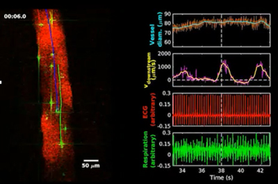
Simultaneous recording of lymph flow speed with ECG and respiration in superficial cervical lymph vessels. Two-photon microscopy imaging (left) and synchronized physiological measurements (right). Trajectories from particle tracking velocimetry are indicated by colored curves tracking the microspheres.
The role of subarachnoid lymphatic-like membrane (SLYM) in traumatic brain injury
The subarachnoid lymphatic-like membrane (SLYM) is a fourth meningeal layer that compartmentalizes the subarachnoid space in the mouse and human brain. The functional characterization of SLYM provides fundamental insights into brain immune barriers and fluid transport. Together with Dr. Maiken Nedergaard, our research tests the interaction between neuroinflammation and SLYM in traumatic brain injury.

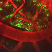
Two-photon microscopy was used to image SLYM (green) and blood vessels (red) after traumatic brain injury in Prox1-EGFP+ reporter mice.
The changes of brain fluid homeostasis in hydrocephalus
Brain fluid homeostasis is the delicate balance of cerebrospinal fluid (CSF) production, circulation, and clearance, which is essential for maintaining normal brain function. When brain fluid homeostasis is disrupted, conditions such as hydrocephalus can develop. Hydrocephalus is characterized by abnormal accumulation of CSF in the brain’s ventricles, leading to increased intracranial pressure and potential damage to brain tissues. It can result from impaired CSF absorption, excessive production, or blockages in CSF pathways. Understanding the mechanisms underlying brain fluid homeostasis and the role of the choroid plexus is critical for developing effective treatments for hydrocephalus and related neurological disorders.

pAAV-CAG-GFP was injected into the lateral ventricle to selectively label the choroid plexus.