Mapping changes in iron distribution and myelin integrity in multiple sclerosis
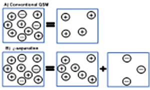
Figure 5a: Schematic illustration of A) conventional QSM – only the sum of susceptibilities within a voxel can be reconstructed, B) -separation – can separate positive and negative susceptibility sources within the same voxel.
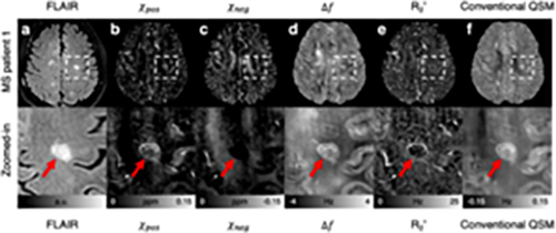
Figure 5b: An MS patient with lesions: a) FLAIR, b) Positive susceptibility (pos) map, c) Negative susceptibility (neg) map, d) Frequency shift (f) map, e) R2' map, f) Conventional QSM map. The zoom-in images show the areas containing typical MS lesions. Red arrows indicate the lesions location in the FLAIR images (adapted from 3).
Multiple sclerosis (MS) is a debilitating disorder characterized by inflammation, demyelination, and neuroaxonal loss within the central nervous system. This condition is often associated with changes in the distribution of iron. Iron is essential for oligodendrocytes, which produce myelin, as well as for cellular energy metabolism and neurotransmitter synthesis. However, an excess iron can be detrimental to cells, leading to oxidative stress that can further damage oligodendrocytes and exacerbate demyelination. Moreover, iron is also involved in mediating the impact of Semaphorin 4A (SEMA 4A) on oligodendrocyte injury. As a result, there exists a complex interaction among oligodendrocytes, iron, and demyelination in the context of MS. Additionally, there is substantial evidence to suggest that all of these factors contribute to cognitive impairment associated with MS.
Our overarching goal of this proposal is to assess the impact iron concentration/ distribution in the pathogenesis of MS on demyelination and axonal injury. To achieve this goal, we intend to conduct a cross-sectional study involving 40 participants, matched for age and sex, consisting of 20 individuals with MS and 20 healthy controls. This study will involve the collection of advanced MRI images, blood markers, and cognitive data. We will address the specific aims listed below. In summary, this cross-sectional study will lay the foundation for future longitudinal studies that intend to monitor inflammations, demyelination and axonal injury in MS. This will be essential in evaluating promising interventions aimed at achieving various outcomes.