Tensor-valued diffusion MRI for neuronal injury in people living with HIV
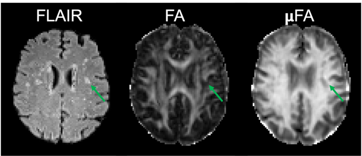
Figure 4: Example images from a 61-year-old HIV+ subject: White matter hyperintensities showing in FLAIR, DTI FA and microscopic fractional anisotropy, μFA quantified using b-tensor encoding.
Globally, 38 million individuals are affected by HIV. While combination antiretroviral treatment (cART) has made HIV manageable, eliminating the virus from the brain remains challenging. Traditional methods for assessing neuroinflammation are invasive or involve radiation, lacking MRI's exceptional spatial resolution.
Diffusion MRI, known for noninvasive detection of brain microstructural abnormalities, faces challenges in regional fiber orientation dispersion. Tensor valued diffusion or b-tensor encoding, employing multiple tensor encodings, addresses these challenges, enabling sub-voxel assessment of axonal integrity through microscopic fractional anisotropy (µFA) and diffusional variance decomposition (MKi and MKa). Our hypothesis is that b-tensor encoding detects subtle changes in gray and white matter microstructural integrity, informing mild chronic neuroinflammation in people living with HIV (PLWH) on cART. We will further improve this technique for faster and high acquisition through the physics informed machine learning approach.
We also aim to explore relationships between b-tensor encoding metrics, extracellular free water, plasma levels of neurofilament light chain (NFL), and glial fibrillary acidic protein (GFAP). Ultimately, we seek to demonstrate that inflammation and neuronal damage correlate with diminished cognitive performance. This study may shed light on the evolution of HIV-associated chronic inflammation and axonal injury, offering sensitivity for evaluating interventions targeting neuroinflammation and other causes of neurodegeneration.