What is an Echocardiogram?
What is an Echocardiogram?
An echocardiogram, also called an "echo," uses sound waves to see the heart's structures and function. It is also called a cardiac ultrasound. The "echo" is used to diagnose and assess problems with the heart.
 Why is an Echocardiogram Performed?
Your child’s doctor may order an echocardiogram to look at the structures and function of your child’s heart. An echocardiogram can show:
- How well the heart is pumping blood
- Exact size and shape of the heart chambers
- Structural or functional problems with the heart valves, such as leaking
- Images of the blood vessels near the heart
- Any blood clots inside the heart
- Abnormal holes in the heart
What to Expect During an Echocardiogram
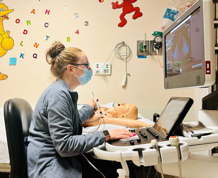
- Before an echocardiogram, a sonographer will attach small plastic adhesive patches (electrodes) to your child's chest to monitor the heart’s rhythm. The echo exam should be painless. The sonographer will place gel on your child's chest and on the transducer.
Your child might feel a slight pressure as the sonographer moves the transducer around his or her chest to get pictures of the heart.
- Once the echocardiogram is done, the sonographer will wipe the gel from your child's chest and remove the electrode patches.
- It is helpful for children to be as still as possible during the echo procedure. If your child is not able to remain still, sedation might be required for the echocardiogram.
The Different Types of Echocardiograms
- Transthoracic (standard) echocardiogram is done by taking pictures using a small camera on the chest while the child is lying on a bed.
- Stress echocardiogram is used to examine what happens to the heart during a period of stress, produced either by medications or by exercise.
- Transesophageal echocardiogram (TEE) is done by placing the camera inside the esophagus. The child is sedated for the TEE, which provides higher-resolution images than a standard.