Gross Photo: Rectal Mass
Olalekan Lanipekun MBBS, Aaron R. Huber, D.O, Roula Katerji MD
Clinical History
An otherwise healthy middle-aged patient presented for screening colonoscopy. The patient was healthy, asymptomatic, and had no family history of colorectal cancer or inflammatory bowel disease.
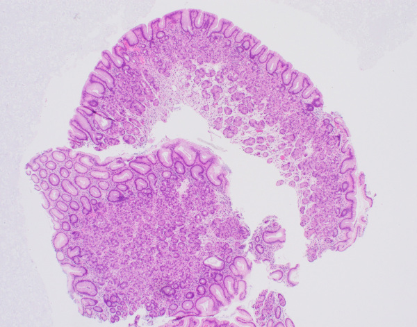
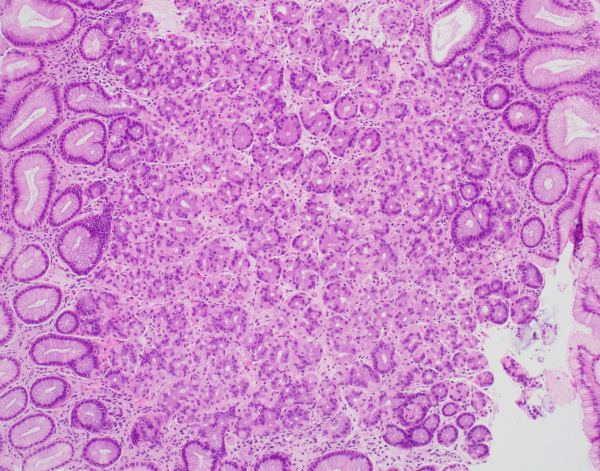
Screening colonoscopy showed a 2.5 cm friable lesion in the distal rectum adjacent to the anal verge. The lesion could not be safely removed during the procedure; however, biopsies were taken. H&E-stained sections of the rectal mucosa demonstrated tightly packed glands with parietal and chief cells resembling oxyntic-type gastric mucosa. No colonic mucosa was identified and no dysplasia or malignancy was seen. (Figures 1-2).
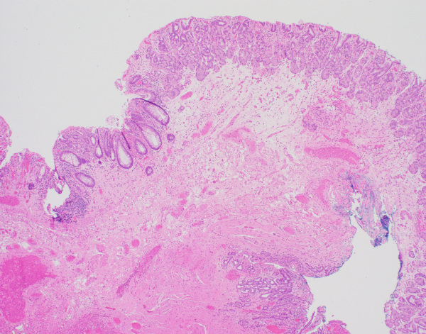
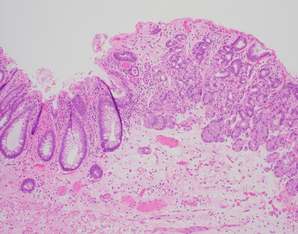
Due to a high clinical suspicion for malignancy endoscopic mucosal resection of the lesion was performed. H&E-stained sections again demonstrated colonic mucosa intermingled with oxyntic-type gastric mucosa. (Figures 3-4). There was no evidence of dysplasia or malignancy.
What is your diagnosis and gross differential diagnosis?
Click image to enlarge.


