Diagnosis
Diagnosis
Serous Cystadenoma
Gross Differential Diagnosis and Discussion
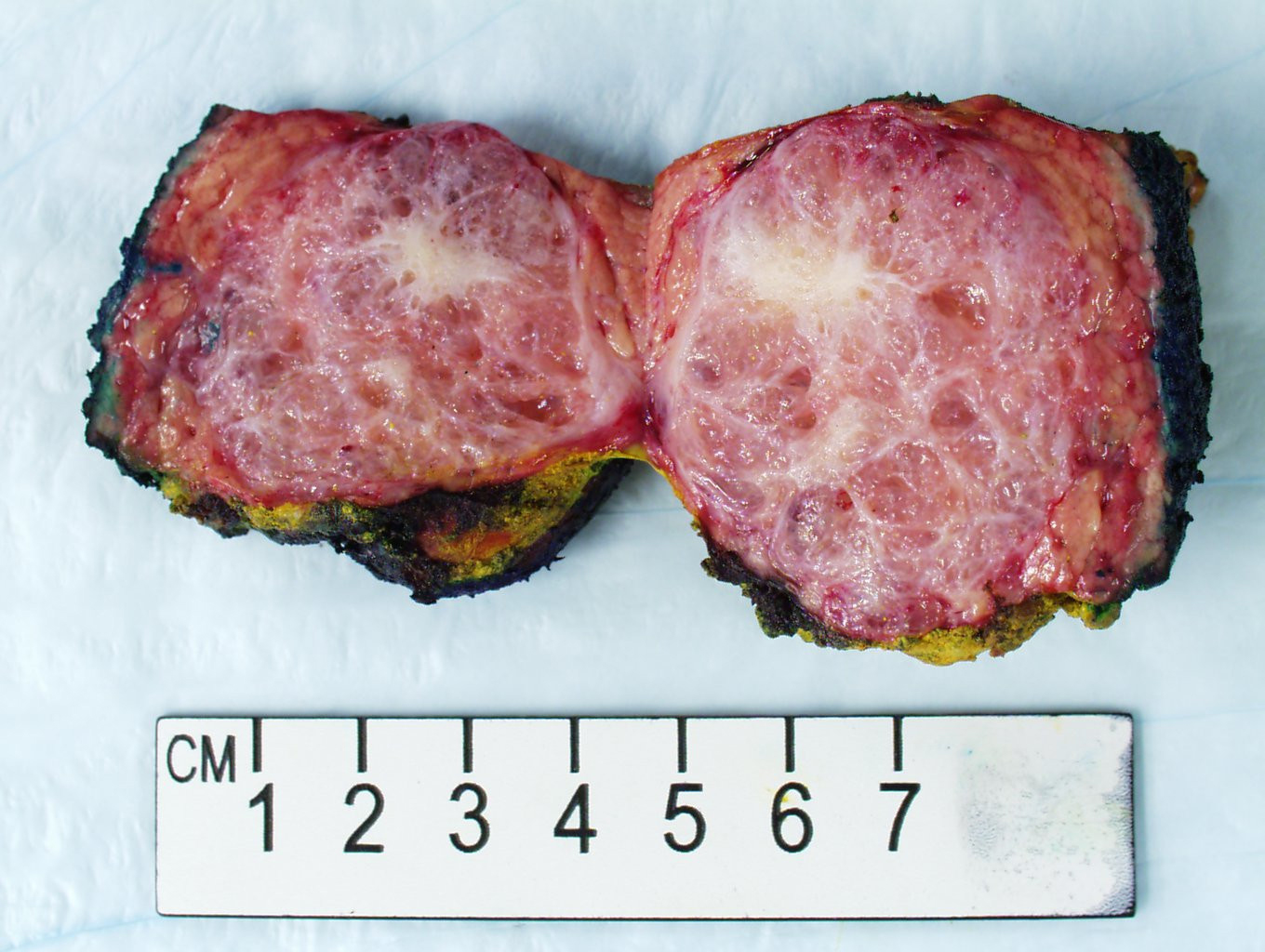
The gross photo demonstrates a well-circumscribed, tan-white, sponge-like mass with a central stellate scar within the pancreatic parenchyma. Serous cystadenoma (SCA) is a benign pancreatic cystic neoplasm composed of cysts lined by bland cuboidal cells with clear, glycogen-rich cytoplasm. Macroscopically, SCAs are well-circumscribed cystic masses with a mean size of 4 cm (range: 1-25 cm). The microcystic variant classically has a sponge-like or honeycomb cut surface. These tumors demonstrate a central scar in approximately 30% of cases.
The macroscopic differential diagnosis includes other pancreatic cysts. Mucinous cystic neoplasm (MCN) is a mucinous cystic epithelial neoplasm of the pancreas with ovarian-type stroma underneath mucinous epithelium. MCNs occur in the body or tail of the pancreas (over 98%), are usually solitary, average 6 cm in size, may be unilocular or multilocular, do not communicate with the pancreatic ductal system, and have thick cyst walls (over 3 mm in thickness). Intraductal papillary mucinous neoplasms (IPMNs) of the pancreas are also composed of cysts lined by mucinous epithelium typically with papillary projections. IPMNs must be grossly visible cystic neoplasms within the main or branch ducts. These tumors produce unilocular or multilocular cysts and connect to the pancreatic ductal system, which is an important differentiating feature. Pancreatic pseudocysts are not true cysts, as the name implies, and are mostly associated with pancreatitis. The "cyst" is usually present in the peripancreatic tissues and may contain necrotic debris or blood. Pancreatic neuroendocrine tumors (Pan-NETs) may have necrosis, hemorrhage, or cystic changes. Pan-NETs are also commonly well-circumscribed, have a capsule, and occur in the tail of the pancreas. Solid pseudopapillary neoplasm (SPN) is a low-grade malignant tumor that nearly uniformly occurs in young women in their 20s (90%). SPNs are large, well-circumscribed and have both solid and cystic components The cysts represent degenerative changes in the neoplasm such as hemorrhage and/or necrosis.
This gross photo of the month concludes our multi-organ macroscopic central scar differential diagnosis which includes:
1. Renal Oncocytoma (50% have a central scar)
https://www.urmc.rochester.edu/pathology-labs/education/pathology-now/gross-photo-case-4.aspx
2. Focal Nodular Hyperplasia (60% have a central scar)
https://www.urmc.rochester.edu/pathology-labs/education/pathology-now/gross-photo-case-3.aspx
3. Pancreatic Serous Cystadenoma (30% have a central scar)
Another example of pancreatic serous cystadenoma with, perhaps, an even better central stellate scar [photo courtesy of Elizabeth Sharratt, PA (ASCP)] (Figure 2).
4. Fibrolamellar Hepatocellular Carcinoma (70% have a central scar)
References
Susan C. Lester, Manual of Surgical Pathology, 3rd Edition. W.B. Saunders, 2010.
Chen W, Ahmed N, Krishna SG. Pancreatic cystic lesions: a focused review on cyst clinicopathological features and advanced diagnosis. Diagnostics. 2023;13:65.
