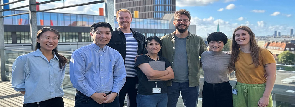
Core Contributors

Construction of viral vectors suitable for in vivo imaging of brain fluid dynamics
I’m involved in molecular work for the production of adeno-associated viral vectors. I’m excited to see how molecular genetics can be applied to color body fluids and to learn how brain fluid circulation/dynamics affects brain states and the associated neuronal activities.
Techniques: Molecular cloning, PCR, DNA design, Fluorescence microscopy, Cell culture, AAV production
Long-term visualization of cerebral blood flow in mice
We are developing a molecular genetics method for the long-term visualization of blood using secretory fluorescent proteins. Our strategy is to target the liver for the blood plasma label production. To this end, we find that AAVs with the AAV8 capsid show good tropism to the liver. Our AAVs can be administered i.v.or i.p.
Techniques: Blood micro-sampling, Fluorescence plate reader analysis, Viral vector inoculation (local and systemic), In vivo two-photon imaging, In vivo one-photon macroscopic imaging
Albumin visualization, astrocytic activity, and vascular dynamics
My research is about how astrocytes modulate brain activity, possibly via vascular dynamics. We are developing a molecular genetics approach to tag albumin with a fluorescent protein, with which we can visualize various extracellular structures in vivo.
Techniques: DNA construction design, Molecular cloning, Primer design for genomic qPCR, In vivo two-photon imaging, In vivo one-photon macroscopic imaging, Optogenetic stimulation, Image analysis for vessel morphology and biosensor readout
Interstitial fluid labeling via secretory fluorescence protein
I am currently engaged in a project to understand astrocytic activity during neuromodulation. It will be wonderful if we can quantitate the extracellular space changes by neuronal or astrocytic activity.
Techniques: Optogenetic stimulation, Viral vector inoculation (local and systemic), In vivo two-photon imaging, Fluorescence signal analysis, Immunohistochemistry
Address:
University of Copenhagen
Faculty of Health and Medical Sciences
Faculty Service, Finance
Blegdamsvej3B
2200 Copenhagen N, Denmark

